


Search Results
Results for: 'cranial fossa'
Cavernous Sinus Larynx Middle Ear Orbit: Granulesm Animation
By: HWC, Views: 10343
The cavernous sinuses are located within the middle cranial fossa, on either side of the sella turcica of the sphenoid bone (which contains the pituitary gland). The cavernous sinuses, a rich plexuses of veins that surround the internal carotid arteries, lie lateral to the pituitary fossa. Ant...
By: Administrator, Views: 715
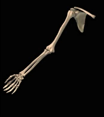
The humerus is a long bone in the arm that runs from the shoulder to the elbow. It connects the scapula and the two bones of the lower arm, the radius and ulna, and consists of three sections. The humeral upper extremity consists of a rounded head, a narrow neck, and two short processes (tubercle...
Central Nervous System Animation
By: Administrator, Views: 14194
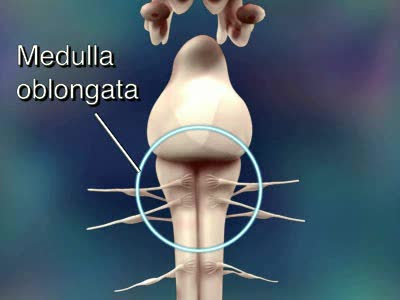
Consists of the brain and spinal cord. CNS receives impulses from throughout the body processes the information responds with an appropriate action Brain and spinal cord can be divided into: gray matter (unsheathed cell bodies and true dendrites) white matter (myelinated nerve fibers) ...
By: Administrator, Views: 15532
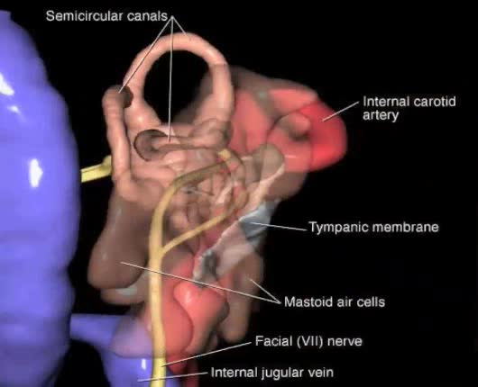
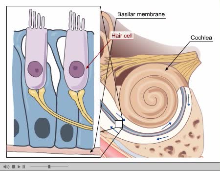
The ear is generally described as having three distinct divisions, each with distinct functions: External ear Middle ear Inner ear The ear contains structures for both the sense of hearing and the sense of balance. Eighth cranial nerve: Also called the acoustic or auditory nerve. Carries...
By: Administrator, Views: 14632
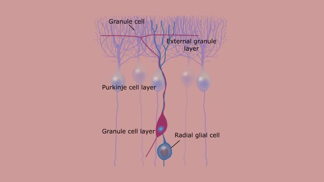
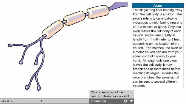
There are several types of neurons, three of which are: Motor neurons, Sensory neurons, Interneurons. The nervous system is usually described as having two interconnected divisions: the central nervous system (CNS) and the peripheral nervous system (PNS). CNS: Includes the brain and spinal...
By: Administrator, Views: 14370
Process of Hearing Sound waves are directed to the eardrum, causing it to vibrate. These vibrations move the three small bones of the middle ear (malleus, incus, and stapes). Movement of stapes at oval window sets up pressure waves in the perilymph and endolymph. Process of Hearing The wav...
By: Administrator, Views: 14393
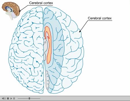
The cerebral cortex (plural cortices), also known as the cerebral mantle, is the outer layer of neural tissue of the cerebrum of the brain, in humans and other mammals. It is separated into two cortices, by the longitudinal fissure that divides the cerebrum into the left and right cerebral hemisp...
Autonomic Nervous System Animation
By: Administrator, Views: 14309
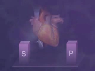
Parasympathetic Division Works to conserve energy and innervate the digestive system. When activated, it: stimulates the salivary and digestive glands. decreases the metabolic rate. slows the heart rate. reduces blood pressure. promotes the passage of material through the intestines along...

Components of the Nervous System
By: Administrator, Views: 539
The nervous system is the part of an animal that coordinates its actions by transmitting signals to and from different parts of its body. The nervous system detects environmental changes that impact the body, then works in tandem with the endocrine system to respond to such events. Nervous tissue...
Advertisement